

Radiotherapy
Radiotherapy is one of the most effective and widely used methods in cancer treatment. At Sante Oncology Centers, we use advanced technologies such as Linear Accelerators (LINAC), Gamma Knife radiosurgery, and brachytherapy to deliver highly precise and personalized cancer treatment.
Our approach is based on patient safety, treatment accuracy, and international clinical standards, supported by a multidisciplinary team of specialists.
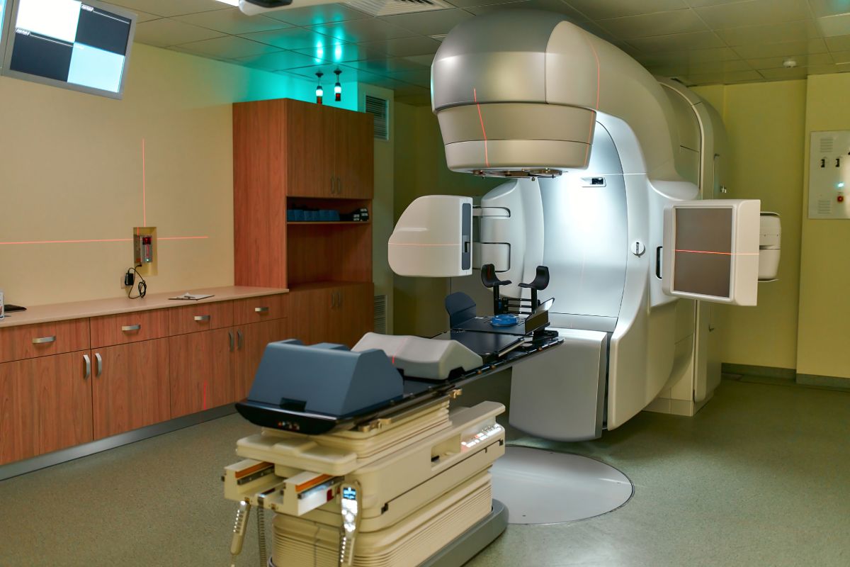
Linear Accelerator (LINAC)
What is a Linac
A Linear Accelerator (LINAC) is an advanced medical device used to deliver high-energy X-ray radiation to cancerous tumors with high precision. A Linear Accelerator (LINAC) is an advanced medical device used to deliver high-energy radiation to tumors with high precision while minimizing exposure to surrounding healthy tissues.
Modern LINAC systems use advanced imaging, computer-assisted planning, and precise patient positioning to ensure accurate dose delivery. Treatments are carefully planned by a multidisciplinary team, including radiation oncologists, medical physicists, and radiotherapy technologists.
LINAC treatments are non-invasive, painless, and usually delivered over multiple sessions, depending on the clinical indication.
Radiotherapy Methods Used with LINAC
External Beam Radiotherapy (EBRT)
External Beam Radiotherapy delivers radiation from outside the body, targeting the tumor from multiple angles. This approach allows for uniform dose distribution and protection of nearby organs at risk.
Intensity-Modulated Radiotherapy (IMRT)
IMRT is an advanced form of radiotherapy that modulates the intensity of radiation beams. This allows:
- Higher doses to the tumor
- Lower doses to surrounding normal tissues
- Improved treatment precision for complex tumor shapes
Image-Guided Radiotherapy (IGRT)
IGRT uses imaging, such as CT or X-ray guidance, immediately before or during treatment to verify tumor position. This increases accuracy and allows for safe dose escalation when clinically appropriate.
Stereotactic Radiotherapy (SRT / SBRT)
Stereotactic techniques deliver highly focused radiation in a small number of sessions. They are used for small, well-defined tumors and require advanced imaging and immobilization systems.
Diseases and Conditions Treated with Radiotherapy
Radiotherapy is an essential component in the treatment of many malignant and selected benign conditions. Treatment decisions are always individualized and based on international clinical guidelines.
Common Cancers Treated with Radiotherapy
- Breast cancer
- Prostate cancer
- Lung cancer
- Head and neck cancers
- Brain tumors
- Gynecological cancers (cervical, uterine)
- Gastrointestinal cancers (rectal, esophageal)
- Skin cancers
- Lymphomas
Stereotactic and Specialized Indications
- Brain metastases
- Arteriovenous malformations (AVMs)
- Selected benign brain tumors
- Functional neurological disorders (in selected cases)
Palliative Radiotherapy
Radiotherapy can also be used to:
- Relieve pain
- Control bleeding
- Reduce tumor-related symptoms
- Improve quality of life in advanced disease
Treatment Planning and Patient Safety
Every radiotherapy treatment is:
- Planned using CT-based simulation
- Reviewed by a multidisciplinary tumor board
- Calculated and verified by medical physicists
- Delivered under strict radiation safety and quality assurance protocols
Patient safety, ethical practice, and informed consent are fundamental principles of our radiotherapy services.
Important Note for Patients
Radiotherapy recommendations are individualized for each patient based on the type and stage of the disease, overall health condition, and previous treatments. All treatment decisions are made collaboratively between the patient and the medical team following comprehensive clinical evaluation.
Gamma Knife Radiosurgery
Gamma Knife Radiosurgery is an advanced, non-invasive treatment method used for the precise management of brain tumors and selected neurological conditions. It delivers highly focused radiation beams to the target area while minimizing exposure to surrounding healthy tissue.
This technology offers an effective alternative to traditional brain surgery and is typically performed in a single session.
What is Gamma Knife Radiosurgery?
Gamma Knife is a specialized form of stereotactic radiosurgery used to treat abnormalities within the brain. It delivers a single, high dose of radiation with sub-millimeter accuracy, targeting tumors and other conditions without affecting surrounding healthy tissue.
It is particularly effective for small to medium-sized lesions and is widely used in modern neuro-oncology.
How the Treatment Works
The precision of Gamma Knife treatment is achieved through advanced imaging techniques such as CT and MRI, combined with computer-assisted planning systems. These technologies create a detailed three-dimensional map of the brain, allowing physicians to accurately identify and target the treatment area.
Multiple radiation beams are directed simultaneously at the target, delivering a high dose to the lesion while protecting surrounding healthy tissue.
Conditions Treated
- Brain tumors
- Brain metastases
- Arteriovenous malformations (AVMs)
- Trigeminal neuralgia
- Acoustic neuroma
- Pituitary tumors
- Meningiomas
- Gliomas
- Epilepsy (selected cases)
- Essential tremor
Treatment Procedure
The procedure begins with the application of a stereotactic frame to ensure precise targeting. Advanced imaging is then performed to identify the exact location of the condition.
During the treatment, the patient lies comfortably while the system delivers multiple focused radiation beams to the target area. The procedure is painless and does not require general anesthesia.
Treatment duration varies depending on the diagnosis, but most sessions are completed within a few hours.
Recovery and Aftercare
Gamma Knife treatment is typically performed on an outpatient basis, allowing patients to return home the same day. Most individuals resume normal activities within a short time.
Some patients may experience mild side effects such as headache or fatigue, which can be managed with medication. Follow-up appointments are scheduled to monitor treatment outcomes.
Brachytherapy (Internal Radiotherapy)
Brachytherapy is a form of internal radiotherapy in which a radioactive source is placed directly inside or very close to the tumor. This technique allows a high dose of radiation to be delivered to the target area while minimizing exposure to surrounding healthy tissues.
Brachytherapy offers highly precise local treatment and may be used alone or in combination with external beam radiotherapy depending on the clinical indication.
What is Brachytherapy?
Brachytherapy delivers radiation internally with high precision, allowing effective treatment of selected tumors while protecting surrounding healthy tissues. Depending on the treatment plan, it may be used alone or combined with other radiotherapy techniques.
Types of Brachytherapy
- High-Dose-Rate (HDR) Brachytherapy
Delivers radiation over a short period using a remotely controlled radioactive source. Usually completed in one or several sessions without prolonged hospitalization. - Low-Dose-Rate (LDR) Brachytherapy
Provides continuous radiation over a longer period, sometimes requiring temporary hospitalization.
Treatment Procedure
1. Comprehensive clinical evaluation and imaging (CT, MRI, or ultrasound)
2. Individualized treatment planning
3. Precise placement of applicators or catheters
4. Radiation dose calculation and verification
5. Safe delivery under continuous monitoring
All procedures are conducted according to international radiotherapy standards and national radiation safety regulations, ensuring patient comfort and safety.
Conditions Treated with Brachytherapy
- Cervical cancer
- Endometrial (uterine) cancer
- Vaginal cancer
Benefits of Brachytherapy
- High radiation dose directly to the tumor
- Reduced exposure to surrounding organs
- Shorter treatment duration
- High precision
- Proven effectiveness in selected cancers
Patient Safety and Quality Assurance
Brachytherapy treatments are:
– Planned using advanced imaging techniques
– Calculated and reviewed by medical physicists
– Delivered under strict radiation protection measures
– Performed with full patient information and consent
Patient comfort, safety, and ethical medical practice are central to all procedures.
Important Information for Patients
The suitability of brachytherapy is determined after a comprehensive clinical evaluation and review by a multidisciplinary tumor board. Final treatment decisions are individualized and discussed in detail with each patient to ensure the safest and most effective approach.
List Of Options
Routine hematology and coagulation testing is available 24 hours a day, 7 days a week. Additionally, advanced hematological tests are a performed using a variety of techniques including flow cytometry and electrophoresis based methods and are available from Monday to Friday, 7 am – 5 pm.


Service FAQ
What is radiotherapy?
Radiotherapy is a treatment method that uses high-energy radiation to destroy cancer cells while protecting surrounding healthy tissues.
Is radiotherapy painful?
No, radiotherapy is completely painless. Patients do not feel the radiation during treatment sessions.
How long does radiotherapy treatment take?
Treatment duration varies depending on the diagnosis, but most sessions last only a few minutes and are repeated over several days or weeks.
What technologies are used in radiotherapy?
Modern radiotherapy uses advanced technologies such as Linear Accelerators (LINAC), IMRT, IGRT, and stereotactic techniques for precise treatment.
What types of cancer can be treated with radiotherapy?
Radiotherapy is commonly used for breast, prostate, lung, brain, and gastrointestinal cancers, among others.
How is patient safety ensured during treatment?
All treatments are carefully planned using imaging technologies and are monitored under strict safety protocols by a specialized team.